Combating Pes Planus
Overview

Also known as fallen arches, the condition of flat feet is characterized by a lack of appropriate arch in the inner foot. It can be a genetic condition or the result of improper body mechanics. Often the whole of the foot will contact the ground. Because a healthy foot is structurally able to support the weight of the body thanks to the bone structure that comprises the arch, a flat foot often is unable to properly support this weight and will cause extreme pressure in the joints above, such as the ankles, knees and hips.
Causes
There are a number of different causes that can lead to flat feet or fallen arches. These include, birth defects, while technically not a defect as such, flat feet can be a normal finding in patients from birth. However, a condition called tarsal coalition may occur where some of the bones in the foot are fused together resulting in a flatfoot. Inflammation or damage of the posterior tibial tendon. This tendon forms the end of a muscle that connects the lower leg to the foot, winding around the ankle and attaching to the inner aspect where the arch is normally present. The main role of the posterior tibial tendon is to invert the foot and maintain the arch height throughout the gait cycle. Torn muscles of the leg and foot can cause flat feet. Problems with the nerve supply to the muscles can result in reduction in tone and fallen arches. Fracture dislocation of the bones in the foot. Severe arthritis. While these are the common causes that can result in fallen arches and flat feet, it is important to recognise that there are certain risk factors that can also lead to this condition. These include advancing age, diabetes mellitus, high blood pressure, obesity and pregnancy.
Symptoms
Some people have fallen arches, and they aren?t even aware of it, fallen arches are sometimes asymptomatic and do not always cause pain. However, for others, the following symptoms may be present. Foot pain, particularly in the arches or heels, leg or back pain, feet feel tired quickly, swelling in the feet and difficulty moving the feet.
Diagnosis
Flat feet are easy to identify while standing or walking. When someone with flat feet stands, their inner foot or arch flattens and their foot may roll over to the inner side. This is known as overpronation. To see whether your foot overpronates, stand on tiptoes or push your big toe back as far as possible. If the arch of your foot doesn't appear, your foot is likely to overpronate when you walk or run. It can be difficult to tell whether a child has flat feet because their arches may not fully develop until they're 10 years of age.
Non Surgical Treatment
The treatment is simple for flat feet. We will carry out a biomechanical assessment and full history, often along side a Computerised Gait Scan to give us an idea of how the foot is compensating. Treatment will be to, control how the foot hits the ground, support the middle of the foot and prevent the arch collapsing, promote normal movement in the front of the foot. The ability to do this will be dictated by the movement within the foot to start with. Treatment for all the above problems are often combined with a physiotherapy session in order to help develop a stretching and strengthening program for the back of the legs and the pelvis in order to allow normal function when the orthoses have been prescribed. If you are born with flat feet you will not grow out of them - if you get orthoses, like glasses you will need them for the rest of your life if you want to correct the mechanics in your foot. In 95% of cases, orthoses will reduce symptoms by at least 85%. In the other 5% we will work with them to get them to this level.
Surgical Treatment

This is rare and usually only offered if patients have significant abnormalities in their bones or muscles. Treatments include joint fusion, reshaping the bones in the foot, and occasionally moving around tendons in the foot to help balance out the stresses (called tendon transfer). Flat feet and fallen arches are common conditions that are in most cases asymptomatic. However, in patients who do have symptoms, treatments are available that can help reduce pain and promote efficient movement. Orthotic devices are well recognised as an excellent treatment and podiatrists can offer these different treatment modalities as individualised treatments for patients.
Also known as fallen arches, the condition of flat feet is characterized by a lack of appropriate arch in the inner foot. It can be a genetic condition or the result of improper body mechanics. Often the whole of the foot will contact the ground. Because a healthy foot is structurally able to support the weight of the body thanks to the bone structure that comprises the arch, a flat foot often is unable to properly support this weight and will cause extreme pressure in the joints above, such as the ankles, knees and hips.
Causes
There are a number of different causes that can lead to flat feet or fallen arches. These include, birth defects, while technically not a defect as such, flat feet can be a normal finding in patients from birth. However, a condition called tarsal coalition may occur where some of the bones in the foot are fused together resulting in a flatfoot. Inflammation or damage of the posterior tibial tendon. This tendon forms the end of a muscle that connects the lower leg to the foot, winding around the ankle and attaching to the inner aspect where the arch is normally present. The main role of the posterior tibial tendon is to invert the foot and maintain the arch height throughout the gait cycle. Torn muscles of the leg and foot can cause flat feet. Problems with the nerve supply to the muscles can result in reduction in tone and fallen arches. Fracture dislocation of the bones in the foot. Severe arthritis. While these are the common causes that can result in fallen arches and flat feet, it is important to recognise that there are certain risk factors that can also lead to this condition. These include advancing age, diabetes mellitus, high blood pressure, obesity and pregnancy.
Symptoms
Some people have fallen arches, and they aren?t even aware of it, fallen arches are sometimes asymptomatic and do not always cause pain. However, for others, the following symptoms may be present. Foot pain, particularly in the arches or heels, leg or back pain, feet feel tired quickly, swelling in the feet and difficulty moving the feet.
Diagnosis
Flat feet are easy to identify while standing or walking. When someone with flat feet stands, their inner foot or arch flattens and their foot may roll over to the inner side. This is known as overpronation. To see whether your foot overpronates, stand on tiptoes or push your big toe back as far as possible. If the arch of your foot doesn't appear, your foot is likely to overpronate when you walk or run. It can be difficult to tell whether a child has flat feet because their arches may not fully develop until they're 10 years of age.
Non Surgical Treatment
The treatment is simple for flat feet. We will carry out a biomechanical assessment and full history, often along side a Computerised Gait Scan to give us an idea of how the foot is compensating. Treatment will be to, control how the foot hits the ground, support the middle of the foot and prevent the arch collapsing, promote normal movement in the front of the foot. The ability to do this will be dictated by the movement within the foot to start with. Treatment for all the above problems are often combined with a physiotherapy session in order to help develop a stretching and strengthening program for the back of the legs and the pelvis in order to allow normal function when the orthoses have been prescribed. If you are born with flat feet you will not grow out of them - if you get orthoses, like glasses you will need them for the rest of your life if you want to correct the mechanics in your foot. In 95% of cases, orthoses will reduce symptoms by at least 85%. In the other 5% we will work with them to get them to this level.
Surgical Treatment
This is rare and usually only offered if patients have significant abnormalities in their bones or muscles. Treatments include joint fusion, reshaping the bones in the foot, and occasionally moving around tendons in the foot to help balance out the stresses (called tendon transfer). Flat feet and fallen arches are common conditions that are in most cases asymptomatic. However, in patients who do have symptoms, treatments are available that can help reduce pain and promote efficient movement. Orthotic devices are well recognised as an excellent treatment and podiatrists can offer these different treatment modalities as individualised treatments for patients.
All The Things You Need To Find Out Regarding Pain In The Arch
Overview
Plantar Fasciitis is an inflammatory process that occurs in the plantar fascia, usually at the insertion to the heel bone (calcaneus). It has been reported that 10% of the worldwide population will suffer from this condition at some point in their lives. The inflammatory process is thought to occur due to repeated trauma to the plantar fascia as it is over-stretched. In other words, the burden on the foot is too great for the plantar fascia to maintain the foot arch and therefore the arch 'collapses' or 'falls' slightly more than it should, thus over-stretching the fascia. This causes damage (micro-tears) in the plantar fascia which triggers the inflammatory response, causing pain. In the vast majority of cases this process occurs at the origin of the plantar fascia at the heel bone.
Causes
Arch pain can be caused by several different factors. Tight calf muscles combined with repeated arch collapse on weight bearing, is the most common cause of arch pain.This can then often lead to a condition called plantar fasciitis, which is an inflammation of the connective tissue that connects the heel with the ball of the foot. When the connective tissue in the bottom of the feet gets stretched out too much, plantar fasciitis can result. The inflammation resulting from plantar fasciitis can result in arch pain. Arch pain caused by plantar fasciitis is often worst after long periods of rest, like when you first wake up in the morning. This is because the plantar fascia tightens when you are off your feet. When you wake up in the morning and start walking around, the plantar fascia stretches out again.
Symptoms
Arch pain symptoms could include any of the following, a dull, constant ache if the ligaments have been stretched, swelling or tenderness in the foot, redness or bruising in the event of a more serious injury, difficulty putting weight on the foot, sharp pain when the foot is turned or manipulated, tenderness when pressure is applied. Because the arch of the foot is such a complex structure, arch pain could be an indicator of several different types of injuries. Chronic illnesses such as arthritis could also cause arch pain, and depending on the cause or source of your pain, you may experience discomfort in a variety of different areas. Ask a doctor if you believe you may have injured your foot arch.
Diagnosis
A patient is asked to step with full body weight on the symptomatic foot, keeping the unaffected foot off the ground. The patient is then instructed to "raise up on the tip toes" of the affected foot. If the posterior tibial tendon has been attenuated or ruptured, the patient will be unable to lift the heel off the floor and rise onto the toes. In less severe cases, the patient will be able to rise on the toes, but the heel will not be noted to invert as it normally does when we rise onto the toes. X-rays can be helpful but are not diagnostic of the adult acquired flatfoot. Both feet, the symptomatic and asymptomatic - will demonstrate a flatfoot deformity on x-ray. Careful observation may show a greater severity of deformity on the affected side.
Non Surgical Treatment
Just as there are many different causes of flat feet, there are also many different treatment options. The most important aspect of treatment is determining the exact type or underlying cause of flat feet that you have. Foot and ankle specialists can determine this through thorough clinical examination and special imaging studies (e.g., x-rays, computed tomography, and/or magnetic resonance imaging). Conservative treatment is effective in the vast majority of flat foot cases, and consists of things such as insoles, splints, manipulation, or casting. Surgery is required much less frequently, and is reserved only for some of the severe types of flat foot that do not respond to conservative therapy.

Surgical Treatment
In rare cases, surgery may be needed if a child has flat feet caused by a problem they're born with (a congenital abnormality). The foot may need to be straightened or the bones may need to be separated if they're fused together. Painkillers and insoles are the first treatment options for flat feet that are caused by a joint problem, such as arthritis or a torn tendon. However, surgery may be recommended if the injury or condition is severely affecting your feet. Where flat feet are caused by a condition that affects the nervous system, special shoes, insoles, or supportive foot or leg braces may be needed. Again, in severe cases, an operation may be needed to straighten the feet.
Stretching Exercises
Below are two simple plantar fasciitis stretching exercises to help improve the flexibility of the muscles and tendons around the foot and ankle. Plantar fasciitis stretch taken from The Stretching Handbook. Kneel on one foot and place your body weight over your knee. Keep your heel on the ground and lean forward. In the photo to the left, the athlete is stretching the arch of her left foot. Kneel on one foot with your hands on the ground. Place your body weight over your knee and slowly move your knee forward. Keep your toes on the ground and arch your foot. In the photo to the right, the athlete is stretching the arch of his right foot.
Plantar Fasciitis is an inflammatory process that occurs in the plantar fascia, usually at the insertion to the heel bone (calcaneus). It has been reported that 10% of the worldwide population will suffer from this condition at some point in their lives. The inflammatory process is thought to occur due to repeated trauma to the plantar fascia as it is over-stretched. In other words, the burden on the foot is too great for the plantar fascia to maintain the foot arch and therefore the arch 'collapses' or 'falls' slightly more than it should, thus over-stretching the fascia. This causes damage (micro-tears) in the plantar fascia which triggers the inflammatory response, causing pain. In the vast majority of cases this process occurs at the origin of the plantar fascia at the heel bone.
Causes
Arch pain can be caused by several different factors. Tight calf muscles combined with repeated arch collapse on weight bearing, is the most common cause of arch pain.This can then often lead to a condition called plantar fasciitis, which is an inflammation of the connective tissue that connects the heel with the ball of the foot. When the connective tissue in the bottom of the feet gets stretched out too much, plantar fasciitis can result. The inflammation resulting from plantar fasciitis can result in arch pain. Arch pain caused by plantar fasciitis is often worst after long periods of rest, like when you first wake up in the morning. This is because the plantar fascia tightens when you are off your feet. When you wake up in the morning and start walking around, the plantar fascia stretches out again.
Symptoms
Arch pain symptoms could include any of the following, a dull, constant ache if the ligaments have been stretched, swelling or tenderness in the foot, redness or bruising in the event of a more serious injury, difficulty putting weight on the foot, sharp pain when the foot is turned or manipulated, tenderness when pressure is applied. Because the arch of the foot is such a complex structure, arch pain could be an indicator of several different types of injuries. Chronic illnesses such as arthritis could also cause arch pain, and depending on the cause or source of your pain, you may experience discomfort in a variety of different areas. Ask a doctor if you believe you may have injured your foot arch.
Diagnosis
A patient is asked to step with full body weight on the symptomatic foot, keeping the unaffected foot off the ground. The patient is then instructed to "raise up on the tip toes" of the affected foot. If the posterior tibial tendon has been attenuated or ruptured, the patient will be unable to lift the heel off the floor and rise onto the toes. In less severe cases, the patient will be able to rise on the toes, but the heel will not be noted to invert as it normally does when we rise onto the toes. X-rays can be helpful but are not diagnostic of the adult acquired flatfoot. Both feet, the symptomatic and asymptomatic - will demonstrate a flatfoot deformity on x-ray. Careful observation may show a greater severity of deformity on the affected side.
Non Surgical Treatment
Just as there are many different causes of flat feet, there are also many different treatment options. The most important aspect of treatment is determining the exact type or underlying cause of flat feet that you have. Foot and ankle specialists can determine this through thorough clinical examination and special imaging studies (e.g., x-rays, computed tomography, and/or magnetic resonance imaging). Conservative treatment is effective in the vast majority of flat foot cases, and consists of things such as insoles, splints, manipulation, or casting. Surgery is required much less frequently, and is reserved only for some of the severe types of flat foot that do not respond to conservative therapy.
Surgical Treatment
In rare cases, surgery may be needed if a child has flat feet caused by a problem they're born with (a congenital abnormality). The foot may need to be straightened or the bones may need to be separated if they're fused together. Painkillers and insoles are the first treatment options for flat feet that are caused by a joint problem, such as arthritis or a torn tendon. However, surgery may be recommended if the injury or condition is severely affecting your feet. Where flat feet are caused by a condition that affects the nervous system, special shoes, insoles, or supportive foot or leg braces may be needed. Again, in severe cases, an operation may be needed to straighten the feet.
Stretching Exercises
Below are two simple plantar fasciitis stretching exercises to help improve the flexibility of the muscles and tendons around the foot and ankle. Plantar fasciitis stretch taken from The Stretching Handbook. Kneel on one foot and place your body weight over your knee. Keep your heel on the ground and lean forward. In the photo to the left, the athlete is stretching the arch of her left foot. Kneel on one foot with your hands on the ground. Place your body weight over your knee and slowly move your knee forward. Keep your toes on the ground and arch your foot. In the photo to the right, the athlete is stretching the arch of his right foot.
Pain In The Arches Cause And Effect
Overview
The sensation of sore, aching feet or arch pain refers to an inflammation and burning sensation at the arch of the foot. This inflammation is due to the excessive stretching of the fibrous tissue located along the bottom surface of the foot, called plantar fascia. Plantar fascitis is the term named after this condition and described as inflammation of the fascia, muscles and ligaments on the bottom of the foot, causing pain in the heel and arch of the foot.

Causes
Tarsal Tunnel Syndrome develops when there is compression on the tibial nerve as it passes through the tarsal tunnel on the inner side of the ankle bone (medial malleolus). It can cause pain on bottom of foot as well as pins and needles. Numbness in the heel can often extend down to the big toe and adjacent three toes. In addition, it may also produce hot and cold sensations along the bottom of the foot. Tarsal Tunnel Syndrome is caused by anything which occupies space in the tarsal tunnel including cysts, ganglions, bone spurs, swelling from ankle injuries or tumours. Treatment aims to reduce the foot arch pain and usually consists of rest, strengthening and stretching exercises, compression bandages and steroid injections. If the pain in bottom of foot persists, surgery may be required.
Symptoms
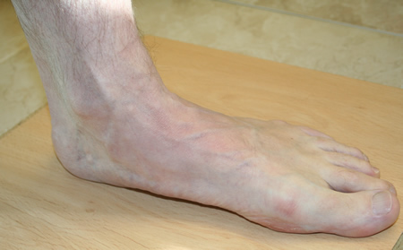
If you've ever seen your footprints in the sand and they looked more like bricks than feet, then you probably have flat feet. Simply stated, a flat foot is a foot that does not have an arch when standing. In the medical world, flat feet are associated with "pronated" feet. Pronated is merely the term used to describe the position of the foot when it is flexed upward (dorsiflexed), turned away from the body (abducted), and the heel is rolled outward (everted), all at the same time. A certain amount of pronation is required for normal walking, but too much pronation is often considered a foot's "worst enemy." Over time, excessive pronation can lead to many unpleasant problems including heel pain, bunions, hammertoes, shin splints, and even knee, hip, or back pain. In fact, one orthopedic surgeon discovered that 95% of his total knee replacement patients and 90% of his total hip replacement patients had flat feet. An easy way to tell if you pronate too much is to take a look at your athletic shoes-excessive wearing of the inside heel (arch side of the shoe) as compared to the outside is a classic indication of excessive pronation.
Diagnosis
After you describe your symptoms and discuss your concerns, your doctor will examine your foot. Your doctor will look for these signs. A high arch. An area of maximum tenderness on the bottom of your foot, just in front of your heel bone. Pain that gets worse when you flex your foot and the doctor pushes on the plantar fascia. The pain improves when you point your toes down. Limited "up" motion of your ankle.
Non Surgical Treatment
Relieving the pain caused by plantar fasciitis boils down to two basic needs. Reduce the inflammation. Support and stretch the plantar fascia. If you can accomplish those two goals, you should note pain relief more quickly. Doctors treating plantar fasciitis will recommend the following options for accomplishing this. Rest, Get off your feet as much as possible when the pain is at its worst. If you must walk or run, try to stay off hard, unforgiving surfaces and wear supporting footwear. Use ice on the arch several times a day to help reduce swelling if necessary. Take Tylenol, Advil, or other over-the-counter pain relievers that contain acetaminophen, ibuprofen, or naproxen to help lessen the inflammation and ease pain. Stretch your toes, calves, and foot repeatedly throughout the day to keep the plantar fasciia limber. Purchase insoles, inserts, or orthopedic shoes designed to support the arch of the foot and wear them at all times. Purchase splints that will stretch the Achilles tendon as you sleep, helping to lessen morning heel pain. If none of the above helps, your doctor may prescribe regular injections of cortisone to control the pain. As a last resort, your doctor may attempt surgery to repair the plantar fascia.

Surgical Treatment
The procedure involves cutting and shifting the bone, and then performing a tendon transfer. First, the surgeon performs a calcaneal osteotomy, cutting the heel bone and shifting it into the correct position. Second, the surgeon transfers the tendon. Reroute the flexor digitorum to replace the troublesome posterior tibial tendon. Finally, the surgeon typically performs one or more fine-tuning procedures that address the patient?s specific foot deformity. Often, the surgeon will lengthen the Achilles tendon because it is common for the mispositioned foot to cause the Achilles to tighten. Occasionally, to increase the arch, the surgeon performs another osteotomy of one of the bones of the midfoot. Occasionally, to point the foot in a straightforward direction, the surgeon performs another osteotomy of the outside portion of the calcaneus.
Prevention
So how do you prevent plantar fasciitis? Factors which can be controlled include training progression, environmental factors, shoes, and strength and flexibility exercises. A useful guideline for a safe training progression is ?the 10% rule.? Limit increases in distance or intensity to 10% a week. For example, if a person is running 60 minutes at a session, 4 times a week, or 240 minutes, she or he can probably increase the running time to 264 minutes (240 + 10%), the following week if all else remains the same. Terrain is also an important factor in training. Running 30 minutes on hills is very different from running 30 minutes on flat surfaces in terms of the forces on the legs and feet. Work up gradually to increase your running time on hills. Also lean forward when running downhill. If you run on a banked or crowned surface, vary the direction you run in so you alternate which leg is higher and which leg is lower on the bank. If you know concrete or asphalt is causing you discomfort, try running on a cinder or composite track. If you are going on vacation and are not used to running on sand or grass, don?t spend your whole vacation doing it.
Stretching Exercises
You may start exercising the muscles of your foot right away by gently stretching and strengthening them. Frozen can roll. Roll your bare injured foot back and forth from your heel to your mid-arch over a frozen juice can. Repeat for 3 to 5 minutes. This exercise is particularly helpful if it is done first thing in the morning. Towel stretch. Sit on a hard surface with your injured leg stretched out in front of you. Loop a towel around your toes and the ball of your foot and pull the towel toward your body keeping your leg straight. Hold this position for 15 to 30 seconds and then relax. Repeat 3 times. Standing calf stretch. Stand facing a wall with your hands on the wall at about eye level. Keep your injured leg back with your heel on the floor. Keep the other leg forward with the knee bent. Turn your back foot slightly inward (as if you were pigeon-toed). Slowly lean into the wall until you feel a stretch in the back of your calf. Hold the stretch for 15 to 30 seconds. Return to the starting position. Repeat 3 times. Do this exercise several times each day. Seated plantar fascia stretch. Sit in a chair and cross the injured foot over the knee of your other leg. Place your fingers over the base of your toes and pull them back toward your shin until you feel a comfortable stretch in the arch of your foot. Hold 15 seconds and repeat 3 times. Plantar fascia massage. Sit in a chair and cross the injured foot over the knee of your other leg. Place your fingers over the base of the toes of your injured foot and pull your toes toward your shin until you feel a stretch in the arch of your foot. With your other hand, massage the bottom of your foot, moving from the heel toward your toes. Do this for 3 to 5 minutes. Start gently. Press harder on the bottom of your foot as you become able to tolerate more pressure.
The sensation of sore, aching feet or arch pain refers to an inflammation and burning sensation at the arch of the foot. This inflammation is due to the excessive stretching of the fibrous tissue located along the bottom surface of the foot, called plantar fascia. Plantar fascitis is the term named after this condition and described as inflammation of the fascia, muscles and ligaments on the bottom of the foot, causing pain in the heel and arch of the foot.
Causes
Tarsal Tunnel Syndrome develops when there is compression on the tibial nerve as it passes through the tarsal tunnel on the inner side of the ankle bone (medial malleolus). It can cause pain on bottom of foot as well as pins and needles. Numbness in the heel can often extend down to the big toe and adjacent three toes. In addition, it may also produce hot and cold sensations along the bottom of the foot. Tarsal Tunnel Syndrome is caused by anything which occupies space in the tarsal tunnel including cysts, ganglions, bone spurs, swelling from ankle injuries or tumours. Treatment aims to reduce the foot arch pain and usually consists of rest, strengthening and stretching exercises, compression bandages and steroid injections. If the pain in bottom of foot persists, surgery may be required.
Symptoms
If you've ever seen your footprints in the sand and they looked more like bricks than feet, then you probably have flat feet. Simply stated, a flat foot is a foot that does not have an arch when standing. In the medical world, flat feet are associated with "pronated" feet. Pronated is merely the term used to describe the position of the foot when it is flexed upward (dorsiflexed), turned away from the body (abducted), and the heel is rolled outward (everted), all at the same time. A certain amount of pronation is required for normal walking, but too much pronation is often considered a foot's "worst enemy." Over time, excessive pronation can lead to many unpleasant problems including heel pain, bunions, hammertoes, shin splints, and even knee, hip, or back pain. In fact, one orthopedic surgeon discovered that 95% of his total knee replacement patients and 90% of his total hip replacement patients had flat feet. An easy way to tell if you pronate too much is to take a look at your athletic shoes-excessive wearing of the inside heel (arch side of the shoe) as compared to the outside is a classic indication of excessive pronation.
Diagnosis
After you describe your symptoms and discuss your concerns, your doctor will examine your foot. Your doctor will look for these signs. A high arch. An area of maximum tenderness on the bottom of your foot, just in front of your heel bone. Pain that gets worse when you flex your foot and the doctor pushes on the plantar fascia. The pain improves when you point your toes down. Limited "up" motion of your ankle.
Non Surgical Treatment
Relieving the pain caused by plantar fasciitis boils down to two basic needs. Reduce the inflammation. Support and stretch the plantar fascia. If you can accomplish those two goals, you should note pain relief more quickly. Doctors treating plantar fasciitis will recommend the following options for accomplishing this. Rest, Get off your feet as much as possible when the pain is at its worst. If you must walk or run, try to stay off hard, unforgiving surfaces and wear supporting footwear. Use ice on the arch several times a day to help reduce swelling if necessary. Take Tylenol, Advil, or other over-the-counter pain relievers that contain acetaminophen, ibuprofen, or naproxen to help lessen the inflammation and ease pain. Stretch your toes, calves, and foot repeatedly throughout the day to keep the plantar fasciia limber. Purchase insoles, inserts, or orthopedic shoes designed to support the arch of the foot and wear them at all times. Purchase splints that will stretch the Achilles tendon as you sleep, helping to lessen morning heel pain. If none of the above helps, your doctor may prescribe regular injections of cortisone to control the pain. As a last resort, your doctor may attempt surgery to repair the plantar fascia.
Surgical Treatment
The procedure involves cutting and shifting the bone, and then performing a tendon transfer. First, the surgeon performs a calcaneal osteotomy, cutting the heel bone and shifting it into the correct position. Second, the surgeon transfers the tendon. Reroute the flexor digitorum to replace the troublesome posterior tibial tendon. Finally, the surgeon typically performs one or more fine-tuning procedures that address the patient?s specific foot deformity. Often, the surgeon will lengthen the Achilles tendon because it is common for the mispositioned foot to cause the Achilles to tighten. Occasionally, to increase the arch, the surgeon performs another osteotomy of one of the bones of the midfoot. Occasionally, to point the foot in a straightforward direction, the surgeon performs another osteotomy of the outside portion of the calcaneus.
Prevention
So how do you prevent plantar fasciitis? Factors which can be controlled include training progression, environmental factors, shoes, and strength and flexibility exercises. A useful guideline for a safe training progression is ?the 10% rule.? Limit increases in distance or intensity to 10% a week. For example, if a person is running 60 minutes at a session, 4 times a week, or 240 minutes, she or he can probably increase the running time to 264 minutes (240 + 10%), the following week if all else remains the same. Terrain is also an important factor in training. Running 30 minutes on hills is very different from running 30 minutes on flat surfaces in terms of the forces on the legs and feet. Work up gradually to increase your running time on hills. Also lean forward when running downhill. If you run on a banked or crowned surface, vary the direction you run in so you alternate which leg is higher and which leg is lower on the bank. If you know concrete or asphalt is causing you discomfort, try running on a cinder or composite track. If you are going on vacation and are not used to running on sand or grass, don?t spend your whole vacation doing it.
Stretching Exercises
You may start exercising the muscles of your foot right away by gently stretching and strengthening them. Frozen can roll. Roll your bare injured foot back and forth from your heel to your mid-arch over a frozen juice can. Repeat for 3 to 5 minutes. This exercise is particularly helpful if it is done first thing in the morning. Towel stretch. Sit on a hard surface with your injured leg stretched out in front of you. Loop a towel around your toes and the ball of your foot and pull the towel toward your body keeping your leg straight. Hold this position for 15 to 30 seconds and then relax. Repeat 3 times. Standing calf stretch. Stand facing a wall with your hands on the wall at about eye level. Keep your injured leg back with your heel on the floor. Keep the other leg forward with the knee bent. Turn your back foot slightly inward (as if you were pigeon-toed). Slowly lean into the wall until you feel a stretch in the back of your calf. Hold the stretch for 15 to 30 seconds. Return to the starting position. Repeat 3 times. Do this exercise several times each day. Seated plantar fascia stretch. Sit in a chair and cross the injured foot over the knee of your other leg. Place your fingers over the base of your toes and pull them back toward your shin until you feel a comfortable stretch in the arch of your foot. Hold 15 seconds and repeat 3 times. Plantar fascia massage. Sit in a chair and cross the injured foot over the knee of your other leg. Place your fingers over the base of the toes of your injured foot and pull your toes toward your shin until you feel a stretch in the arch of your foot. With your other hand, massage the bottom of your foot, moving from the heel toward your toes. Do this for 3 to 5 minutes. Start gently. Press harder on the bottom of your foot as you become able to tolerate more pressure.
Adult Aquired FlatFeet Causes
Overview
Flatfoot may sound being a characteristic of the specific water animal instead of any human problem. Flatfoot can be a condition in which the actual arch with the foot will be fallen as well as the foot will be pointed outward. Throughout distinction to some flatfoot condition which has always been present, this type develops following your skeleton has reached maturity. There are several circumstances which can lead to fallen arches, which includes fracture, dislocation, tendon laceration, tarsal coalition, as well as arthritis. one of the very typical conditions that can lead to this foot problem is posterior tibial tendon dysfunction. the posterior tibial tendon attaches your calf muscle mass to the bones on the inside of the foot and is crucial in holding up as well as supporting the actual arch. An acute injury or perhaps overuse may cause this tendon for you to become inflamed or perhaps torn, and additionally the arch in the foot will slowly fall more than time.

Causes
The most common cause associated with acquired adult flatfoot is actually posterior tibial tendon dysfunction. Exactly what leads to adult acquired flat foot? Fracture or dislocation. Tendon laceration. Tarsal Coalition. Arthritis. Neuroarthropathy. Neurological weakness.
Symptoms
The the signs of PTTD may include pain, swelling, any flattening in the arch, as well as inward rolling in the ankle. While the situation progresses, the particular signs and symptoms will change. Pertaining To example, later, because the arch starts to flatten, there might still be pain on the inside of the foot along with ankle. However at this point, your foot and toes start to flip outward as well as the ankle rolls inward. As PTTD grows more advanced, the particular arch flattens a lot more and the pain usually shifts towards the outside involving the foot, under the particular ankle. Your tendon has deteriorated considerably and arthritis usually develops within the foot. Inside much more severe cases, arthritis might also develop within the ankle. Symptoms, which might appear in several persons with flexible flatfoot, include. Pain within the heel, arch, ankle, or even across the outside the foot. ?Turned-in? ankle. Pain associated with a shin splint. General weakness / fatigue within the foot as well as leg.
Diagnosis
First, each feet needs to be able to be analyzed with most the patient standing and the entire lower extremity visible. Your foot should be inspected coming from higher than as well as via behind your patient, as valgus angulation with the hindfoot is best appreciated when the foot can be viewed through behind. Johnson described the so-called more-toes sign: with increased advanced deformity as well as abduction of the forefoot, much more of the particular lateral toes become noticeable if the foot will be viewed through behind. the single-limb heel-rise test is surely an superb determinant with the operate with the posterior tibial tendon. the patient will be asked to try to rise to the ball of one foot although one other foot is actually suspended off the particular floor. under regular circumstances, the particular posterior tibial muscle, which usually inverts and stabilizes your hindfoot, can be activated since the patient begins to rise on the forefoot. The Particular gastrocnemius-soleus muscle mass group then elevates the actual calcaneus, as well as the heel-rise is actually accomplished. Along With dysfunction of the posterior tibial tendon, however, inversion in the heel can be weak, and both the actual heel remains within valgus or the affected person can be not able to rise onto the forefoot. In the actual event that the patient may do a single-limb heel-rise, the actual limb might become stressed further by simply asking the actual patient to do this maneuver repetitively.
Non surgical Treatment
Initial remedy is situated about the degree of deformity and adaptability in original presentation. Conservative treatment method consists of orthotics or even ankle foot orthoses (AFO) to support the posterior tibial tendon (PT) and additionally the longitudinal arch, anti-inflammatories to help decrease pain as well as inflammation, activity modification which may include immobilization with the foot and also physical therapy to assist strengthen along with rehabilitate the particular tendon.

Surgical Treatment
If conservative remedy doesn't provide relief associated with pain as well as disability then surgery is actually considered. Numerous factors decide if the individual is really a surgical candidate. That They consist of age, obesity, diabetes, vascular status, and the ability to become compliant along with post-operative care. Surgery typically needs a prolonged period of time associated with nonweightbearing immobilization. Total recovery ranges coming from three months to no much less than one year. Clinical, x-ray, along with MRI examination are all used to select the appropriate surgical procedure.
Flatfoot may sound being a characteristic of the specific water animal instead of any human problem. Flatfoot can be a condition in which the actual arch with the foot will be fallen as well as the foot will be pointed outward. Throughout distinction to some flatfoot condition which has always been present, this type develops following your skeleton has reached maturity. There are several circumstances which can lead to fallen arches, which includes fracture, dislocation, tendon laceration, tarsal coalition, as well as arthritis. one of the very typical conditions that can lead to this foot problem is posterior tibial tendon dysfunction. the posterior tibial tendon attaches your calf muscle mass to the bones on the inside of the foot and is crucial in holding up as well as supporting the actual arch. An acute injury or perhaps overuse may cause this tendon for you to become inflamed or perhaps torn, and additionally the arch in the foot will slowly fall more than time.
Causes
The most common cause associated with acquired adult flatfoot is actually posterior tibial tendon dysfunction. Exactly what leads to adult acquired flat foot? Fracture or dislocation. Tendon laceration. Tarsal Coalition. Arthritis. Neuroarthropathy. Neurological weakness.
Symptoms
The the signs of PTTD may include pain, swelling, any flattening in the arch, as well as inward rolling in the ankle. While the situation progresses, the particular signs and symptoms will change. Pertaining To example, later, because the arch starts to flatten, there might still be pain on the inside of the foot along with ankle. However at this point, your foot and toes start to flip outward as well as the ankle rolls inward. As PTTD grows more advanced, the particular arch flattens a lot more and the pain usually shifts towards the outside involving the foot, under the particular ankle. Your tendon has deteriorated considerably and arthritis usually develops within the foot. Inside much more severe cases, arthritis might also develop within the ankle. Symptoms, which might appear in several persons with flexible flatfoot, include. Pain within the heel, arch, ankle, or even across the outside the foot. ?Turned-in? ankle. Pain associated with a shin splint. General weakness / fatigue within the foot as well as leg.
Diagnosis
First, each feet needs to be able to be analyzed with most the patient standing and the entire lower extremity visible. Your foot should be inspected coming from higher than as well as via behind your patient, as valgus angulation with the hindfoot is best appreciated when the foot can be viewed through behind. Johnson described the so-called more-toes sign: with increased advanced deformity as well as abduction of the forefoot, much more of the particular lateral toes become noticeable if the foot will be viewed through behind. the single-limb heel-rise test is surely an superb determinant with the operate with the posterior tibial tendon. the patient will be asked to try to rise to the ball of one foot although one other foot is actually suspended off the particular floor. under regular circumstances, the particular posterior tibial muscle, which usually inverts and stabilizes your hindfoot, can be activated since the patient begins to rise on the forefoot. The Particular gastrocnemius-soleus muscle mass group then elevates the actual calcaneus, as well as the heel-rise is actually accomplished. Along With dysfunction of the posterior tibial tendon, however, inversion in the heel can be weak, and both the actual heel remains within valgus or the affected person can be not able to rise onto the forefoot. In the actual event that the patient may do a single-limb heel-rise, the actual limb might become stressed further by simply asking the actual patient to do this maneuver repetitively.
Non surgical Treatment
Initial remedy is situated about the degree of deformity and adaptability in original presentation. Conservative treatment method consists of orthotics or even ankle foot orthoses (AFO) to support the posterior tibial tendon (PT) and additionally the longitudinal arch, anti-inflammatories to help decrease pain as well as inflammation, activity modification which may include immobilization with the foot and also physical therapy to assist strengthen along with rehabilitate the particular tendon.
Surgical Treatment
If conservative remedy doesn't provide relief associated with pain as well as disability then surgery is actually considered. Numerous factors decide if the individual is really a surgical candidate. That They consist of age, obesity, diabetes, vascular status, and the ability to become compliant along with post-operative care. Surgery typically needs a prolonged period of time associated with nonweightbearing immobilization. Total recovery ranges coming from three months to no much less than one year. Clinical, x-ray, along with MRI examination are all used to select the appropriate surgical procedure.
What Are Usually The Principal Brings About Regarding Adult Aquired Flat Feet ?
Overview
Adult acquired flatfoot is often a complex disorder, along with diverse signs and also symptoms and also varying examples of deformity and also disability. You could find a quantity of kinds of flatfoot, just about all of that have 1 characteristic throughout common-partial or even total collapse (loss) of the arch. Various Other characteristics shared by nearly all forms of flatfoot contain ?Toe drift,? the location where the toes and front part of your foot point outward. Your heel tilts towards the outside and additionally the ankle appears to show in. The brief Achilles tendon or even calf muscle, which in turn causes the particular heel to always be able to lift off the particular floor earlier when strolling and may act as the deforming force. Inside addition, additional deformities such bunions as well as hammertoes may occur and also trigger pain inside people which have flexible flatfoot. Well Being problems like rheumatoid arthritis, diabetes along with obesity could improve your likelihood of creating flatfoot and could (or might not) allow it in order to be more challenging to treat. This specific article provides a short overview of the problems that can lead to AAFD. Further details regarding one of the most typical circumstances that cause an acquired flatfoot in add-on for you to their therapy alternatives are supplied throughout separate articles. Hyperlinks to people content is provided.

Causes
As discussed above, many well being circumstances could create a painful flatfoot. Damage towards the posterior tibial tendon may become the most typical trigger involving AAFD. Your posterior tibial tendon will be among the most essential tendons with the leg. It starts at a muscle mass within the calf, travels on the inside regarding the low leg as well as attaches towards the bones about the inside of the foot. the principal perform regarding this tendon is to keep up the arch and also assistance your foot whenever you walk. In the particular event that your tendon becomes inflamed or torn, the particular arch will slowly collapse. women and individuals over 40 are usually more inclined for you to develop problems with the posterior tibial tendon. Various Other risk aspects include obesity, diabetes, along with hypertension. having flat feet since childhood raises the risk of creating any tear in the posterior tibial tendon. in addition, individuals who're associated with high impact sports, like basketball, tennis, or soccer, may get tears in the tendon coming from repetitive use. Inflammatory arthritis, for example rheumatoid arthritis, could cause a painful flatfoot. This sort regarding arthritis attacks certainly not only the cartilage inside the joints, but additionally the particular ligaments that will offer the foot. Inflammatory arthritis not just leads to pain, but also causes your foot to be able to alter shape and turn into flat. Your arthritis can affect the trunk with the foot as well as the midst of foot, each associated with that may result in the fallen arch.
Symptoms
Symptoms are generally minor and could go unnoticed, Pain dominates, as opposed to deformity. Minor swelling could always be visible over the course of the actual tendon. Pain and swelling along the course of the tendon. visible decline in arch height. Aduction in the forefoot upon rearfoot. Subluxed tali along with navicular joints. Deformation only from that point will be still flexible. Considerable deformity along with weakness. Significant pain. Arthritic changes in the tarsal joints. Deformation only in that point is rigid.
Diagnosis
Posterior Tibial Tendon Dysfunction is actually identified as having cautious clinical observation with the patient?s gait (walking), array of motion screening for the foot and also ankle joints, and diagnostic imaging. people along with flatfoot deformity walk with the heel angled outward, furthermore called over-pronation. Even though it truly is normal for the arch for you to impact the bottom regarding shock absorption, people with PTTD have an arch that completely collapses for the ground along with doesn't reform an arch through the entire gait period. Right After evaluating the particular ambulation pattern, the actual foot along with ankle selection of motion needs in order to be tested. usually the particular affected foot will have decreased motion towards the ankle joint and furthermore the hindfoot. Muscle Mass power could also become weaker as well. An easy test to do pertaining to PTTD is the single heel raise in which the individual will be asked to raise up around the ball associated with his as well as her effected foot. A New regular foot sort can easily lift up around the toes without pain and the heel will invert slightly when anyone offers fully raised the particular heel up through the test. Inside early phases regarding PTTD your individual could always be in a situation to lift up the actual heel however the heel is certainly not going to invert. An elongated or perhaps torn posterior tibial tendon, that is a mid to become able to late discovering involving PTTD, will prohibit the particular patient coming from totally rising up about the heel and can trigger intense pain towards the arch. Finally diagnostic imaging, although utilized by yourself cannot diagnose PTTD, may provide further details for an accurate diagnosis regarding flatfoot deformity. Xrays in the foot can show the practitioner crucial angular relationships of the hindfoot as well as forefoot that aid diagnose flatfoot deformity. Many of the time, an MRI isn't necessary to diagnose PTTD yet is actually a instrument that should always be considered within advanced cases associated with flatfoot deformity. In your event that any partial tear with the posterior tibial tendon is regarding concern, then an MRI may display the anatomic location in the tear and the extensiveness in the injury.
Non surgical Treatment
Nonoperative therapy with regard to posterior tibial tendon dysfunction has been demonstrated to yield 67% good-to-excellent leads to 49 patients with stage 2 as well as 3 deformities. a rigid UCBL orthosis with a medial forefoot submit was used within nonobese patients using versatile heel deformities correctible for you to neutral and also lower than 10? involving forefoot varus. The molded ankle foot orthosis was utilized inside obese patients along with fixed deformity and forefoot varus greater compared to 10?. average duration of orthotic use has been 15 months. four patients ultimately elected to possess surgery. The Particular authors concluded that will orthotic management will be productive within older low-demand patients and also that surgical therapy could be reserved for anyone patients which fail nonoperative treatment.
Surgical Treatment
If conservative therapy doesn't offer relief involving pain along with disability then surgery will be considered. Numerous factors determine whether a individual can end up being a surgical candidate. they contain age, obesity, diabetes, vascular status, as well as the ability to be compliant with post-operative care. Surgery generally requires a protracted period regarding time involving nonweightbearing immobilization. Total recovery ranges through three several weeks to one year. Clinical, x-ray, as well as MRI examination tend to be almost all accustomed to select the right surgical procedure.
Adult acquired flatfoot is often a complex disorder, along with diverse signs and also symptoms and also varying examples of deformity and also disability. You could find a quantity of kinds of flatfoot, just about all of that have 1 characteristic throughout common-partial or even total collapse (loss) of the arch. Various Other characteristics shared by nearly all forms of flatfoot contain ?Toe drift,? the location where the toes and front part of your foot point outward. Your heel tilts towards the outside and additionally the ankle appears to show in. The brief Achilles tendon or even calf muscle, which in turn causes the particular heel to always be able to lift off the particular floor earlier when strolling and may act as the deforming force. Inside addition, additional deformities such bunions as well as hammertoes may occur and also trigger pain inside people which have flexible flatfoot. Well Being problems like rheumatoid arthritis, diabetes along with obesity could improve your likelihood of creating flatfoot and could (or might not) allow it in order to be more challenging to treat. This specific article provides a short overview of the problems that can lead to AAFD. Further details regarding one of the most typical circumstances that cause an acquired flatfoot in add-on for you to their therapy alternatives are supplied throughout separate articles. Hyperlinks to people content is provided.
Causes
As discussed above, many well being circumstances could create a painful flatfoot. Damage towards the posterior tibial tendon may become the most typical trigger involving AAFD. Your posterior tibial tendon will be among the most essential tendons with the leg. It starts at a muscle mass within the calf, travels on the inside regarding the low leg as well as attaches towards the bones about the inside of the foot. the principal perform regarding this tendon is to keep up the arch and also assistance your foot whenever you walk. In the particular event that your tendon becomes inflamed or torn, the particular arch will slowly collapse. women and individuals over 40 are usually more inclined for you to develop problems with the posterior tibial tendon. Various Other risk aspects include obesity, diabetes, along with hypertension. having flat feet since childhood raises the risk of creating any tear in the posterior tibial tendon. in addition, individuals who're associated with high impact sports, like basketball, tennis, or soccer, may get tears in the tendon coming from repetitive use. Inflammatory arthritis, for example rheumatoid arthritis, could cause a painful flatfoot. This sort regarding arthritis attacks certainly not only the cartilage inside the joints, but additionally the particular ligaments that will offer the foot. Inflammatory arthritis not just leads to pain, but also causes your foot to be able to alter shape and turn into flat. Your arthritis can affect the trunk with the foot as well as the midst of foot, each associated with that may result in the fallen arch.
Symptoms
Symptoms are generally minor and could go unnoticed, Pain dominates, as opposed to deformity. Minor swelling could always be visible over the course of the actual tendon. Pain and swelling along the course of the tendon. visible decline in arch height. Aduction in the forefoot upon rearfoot. Subluxed tali along with navicular joints. Deformation only from that point will be still flexible. Considerable deformity along with weakness. Significant pain. Arthritic changes in the tarsal joints. Deformation only in that point is rigid.
Diagnosis
Posterior Tibial Tendon Dysfunction is actually identified as having cautious clinical observation with the patient?s gait (walking), array of motion screening for the foot and also ankle joints, and diagnostic imaging. people along with flatfoot deformity walk with the heel angled outward, furthermore called over-pronation. Even though it truly is normal for the arch for you to impact the bottom regarding shock absorption, people with PTTD have an arch that completely collapses for the ground along with doesn't reform an arch through the entire gait period. Right After evaluating the particular ambulation pattern, the actual foot along with ankle selection of motion needs in order to be tested. usually the particular affected foot will have decreased motion towards the ankle joint and furthermore the hindfoot. Muscle Mass power could also become weaker as well. An easy test to do pertaining to PTTD is the single heel raise in which the individual will be asked to raise up around the ball associated with his as well as her effected foot. A New regular foot sort can easily lift up around the toes without pain and the heel will invert slightly when anyone offers fully raised the particular heel up through the test. Inside early phases regarding PTTD your individual could always be in a situation to lift up the actual heel however the heel is certainly not going to invert. An elongated or perhaps torn posterior tibial tendon, that is a mid to become able to late discovering involving PTTD, will prohibit the particular patient coming from totally rising up about the heel and can trigger intense pain towards the arch. Finally diagnostic imaging, although utilized by yourself cannot diagnose PTTD, may provide further details for an accurate diagnosis regarding flatfoot deformity. Xrays in the foot can show the practitioner crucial angular relationships of the hindfoot as well as forefoot that aid diagnose flatfoot deformity. Many of the time, an MRI isn't necessary to diagnose PTTD yet is actually a instrument that should always be considered within advanced cases associated with flatfoot deformity. In your event that any partial tear with the posterior tibial tendon is regarding concern, then an MRI may display the anatomic location in the tear and the extensiveness in the injury.
Non surgical Treatment
Nonoperative therapy with regard to posterior tibial tendon dysfunction has been demonstrated to yield 67% good-to-excellent leads to 49 patients with stage 2 as well as 3 deformities. a rigid UCBL orthosis with a medial forefoot submit was used within nonobese patients using versatile heel deformities correctible for you to neutral and also lower than 10? involving forefoot varus. The molded ankle foot orthosis was utilized inside obese patients along with fixed deformity and forefoot varus greater compared to 10?. average duration of orthotic use has been 15 months. four patients ultimately elected to possess surgery. The Particular authors concluded that will orthotic management will be productive within older low-demand patients and also that surgical therapy could be reserved for anyone patients which fail nonoperative treatment.
Surgical Treatment
If conservative therapy doesn't offer relief involving pain along with disability then surgery will be considered. Numerous factors determine whether a individual can end up being a surgical candidate. they contain age, obesity, diabetes, vascular status, as well as the ability to be compliant with post-operative care. Surgery generally requires a protracted period regarding time involving nonweightbearing immobilization. Total recovery ranges through three several weeks to one year. Clinical, x-ray, as well as MRI examination tend to be almost all accustomed to select the right surgical procedure.